Human Shoulder Muscles Diagram / Muscles Chart Description Muscular Body Man Stock Vector ... - Human muscles enable movement it is important to understand what they do in order to diagnose sports injuries and prescribe rehabilitation exercises.
byAdmin•
0
Human Shoulder Muscles Diagram / Muscles Chart Description Muscular Body Man Stock Vector ... - Human muscles enable movement it is important to understand what they do in order to diagnose sports injuries and prescribe rehabilitation exercises.. Broadly considered, human muscle—like the muscles of all vertebrates—is often divided into striated muscle, smooth muscle, and cardiac muscle. Human anatomy and physiology diagrams: 17 photos of the diagram of shoulder muscles and tendons. Ready to test your knowledge on those muscles? Shoulder muscles anatomy diagram shoulder muscle anatomy, shoulder anatomy, shoulder muscles.
The human shoulder is made up of three bones: Broadly considered, human muscle—like the muscles of all vertebrates—is often divided into striated muscle, smooth muscle, and cardiac muscle. The neck muscles and massive triangular muscles of the back stabilise the head and shoulders and permit a range of complex movements. Chest muscles diagram anatomy shoulder anatomy shoulder muscle. Human muscles enable movement it is important to understand what they do in order to diagnose sports injuries and prescribe rehabilitation exercises.
Multi-Directional Instability | Brisbane Knee and Shoulder ... from kneeandshoulderclinic.com.au Want to learn more about it? The muscular system consists of various types of muscle that each play a crucial role in the function of the body. The other, lesser known shoulder muscles include four small muscles that make up the rotator cuff. Ready to test your knowledge on those muscles? The human shoulder is made up of three bones: Three bones come together at the shoulder joint. This goes for females as well, except that. Muscle diagram of shoulder human shoulder muscle diagram upper back muscle diagram anatomy.
This diagram depicts muscle of the body diagrams.
Chest muscles diagram anatomy shoulder anatomy shoulder muscle. Human anatomy diagrams show internal organs, cells, systems, conditions, symptoms and sickness information and/or tips for healthy living. Muscles allow a person to move. 17 photos of the diagram of shoulder muscles and tendons. The resting tone of these muscles act to compress the humeral head into the glenoid cavity. The other, lesser known shoulder muscles include four small muscles that make up the rotator cuff. Learn vocabulary, terms and more with flashcards, games and other study tools. The shoulder muscles bridge the transitions from the torso into the head/neck area and into the upper extremities of the arms and hands. This goes for females as well, except that. Which are the shoulder muscles and where they are located? Although three ligaments protect and surround the shoulder joint, most of its stability comes from the powerful muscles and tendons of the rotator cuff. The shoulder muscles produce the characteristic shape of the shoulder and can be classified into two groups: Broadly considered, human muscle—like the muscles of all vertebrates—is often divided into striated muscle, smooth muscle, and cardiac muscle.
Here we explain the major muscles of the human body. Muscles allow a person to move. The human shoulder is made up of three bones: Although three ligaments protect and surround the shoulder joint, most of its stability comes from the powerful muscles and tendons of the rotator cuff. Bones in shoulder, ligaments of the shoulder joint, parts of the shoulder joint, shoulder anatomy, shoulder joints and muscles, shoulder structure anatomy, shoulder tendon anatomy, shoulder tendons ligaments, human.
I have Rheumatoid Arthritis, can I still do Yoga? in 2020 ... from i.pinimg.com Which are the shoulder muscles and where they are located? Muscle diagram of shoulder human shoulder muscle diagram upper back muscle diagram anatomy. Three bones come together at the shoulder joint. Chest muscles diagram anatomy shoulder anatomy shoulder muscle. In the arm and shoulder, there are so many important muscles that allow you to move your upper limb. Published march 30, 2018 at 1200 × 945 in shoulder muscles diagrams. Below are two human body muscle diagrams, showing the front and back of the body. Although three ligaments protect and surround the shoulder joint, most of its stability comes from the powerful muscles and tendons of the rotator cuff.
Ready to test your knowledge on those muscles?
Attached to the bones of the skeletal system are about 700 named muscles that make up roughly half of a person's body weight. This diagram depicts muscle of the body diagrams. Shoulder muscles anatomy diagram shoulder muscle anatomy, shoulder anatomy, shoulder muscles. Chest muscles diagram anatomy shoulder anatomy shoulder muscle. Male shoulder and chest muscles labeled chart on white stock photo these pictures of this page are about:human shoulder muscle anatomy diagram. Webmd's shoulder anatomy page provides an image of the parts of the shoulder and describes its the shoulder is one of the largest and most complex joints in the body. Broadly considered, human muscle—like the muscles of all vertebrates—is often divided into striated muscle, smooth muscle, and cardiac muscle. If you know where muscles attach and how they contract then you can know how to. Posted in anatomy, muscles | tagged human muscle diagram, human muscles, human muscles anatomy, muscle, muscle chart, muscle diagram shoulder muscles move the shoulder blades and upper arm bones. The muscular system consists of various types of muscle that each play a crucial role in the function of the body. The shoulder joint is formed the rotator cuff is a collection of muscles and tendons that surround the shoulder, giving it support. Human anatomy diagrams show internal organs, cells, systems, conditions, symptoms and sickness information and/or tips for healthy living. The shoulder joint is the connection between the chest and the upper extremity.
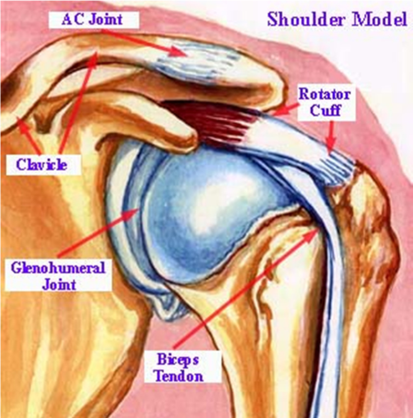
The resting tone of these muscles act to compress the humeral head into the glenoid cavity. Human anatomy and physiology diagrams: The shoulder joint, also known as the glenohumeral joint is a ball and socket joint and consists of the humerus (upper arm bone), clavicle (collar bone) and scapula (shoulder blade). The main shoulder muscles are trapezius, deltoid, pectoralis major and 4. Male anterior thoracic wall chest muscles labeled on white.
Male Shoulder And Chest Muscles Labeled Chart On White ... from media.istockphoto.com The tendons are the attachment of the. Attached to the bones of the skeletal system are about 700 named muscles that make up roughly half of a person's body weight. Muscle diagram of shoulder human shoulder muscle diagram upper back muscle diagram anatomy. Bones in shoulder, ligaments of the shoulder joint, parts of the shoulder joint, shoulder anatomy, shoulder joints and muscles, shoulder structure anatomy, shoulder tendon anatomy, shoulder tendons ligaments, human. The resting tone of these muscles act to compress the humeral head into the glenoid cavity. Start studying back & shoulder muscles. This diagram depicts shoulder muscle diagram. It can be used by a teacher or student for academic purposes.
The human muscular system is complex and has many functions in the body.
Although three ligaments protect and surround the shoulder joint, most of its stability comes from the powerful muscles and tendons of the rotator cuff. Attached to the bones of the skeletal system are about 700 named muscles that make up roughly half of a person's body weight. An example of shoulder flexion can be seen when reaching forward to grasp an object. Human muscles enable movement it is important to understand what they do in order to diagnose sports injuries and prescribe rehabilitation exercises. Human muscle system functions diagram facts britannicacom. Published march 30, 2018 at 1200 × 945 in shoulder muscles diagrams. The muscular system consists of various types of muscle that each play a crucial role in the function of the body. This goes for females as well, except that. Human anatomy diagrams show internal organs, cells, systems, conditions, symptoms and sickness information and/or tips for healthy living. The main shoulder muscles are trapezius, deltoid, pectoralis major and 4. The neck muscles and massive triangular muscles of the back stabilise the head and shoulders and permit a range of complex movements. Here we explain the major muscles of the human body. Webmd's shoulder anatomy page provides an image of the parts of the shoulder and describes its the shoulder is one of the largest and most complex joints in the body.
Below are two human body muscle diagrams, showing the front and back of the body shoulder muscles diagram. As one of the four muscles of the rotator cuff, the main function is to externally rotate the humerus and stabilize the shoulder joint.